Skin Cancer Clinic Mount Martha

About Skin Cancer

Cryotherapy

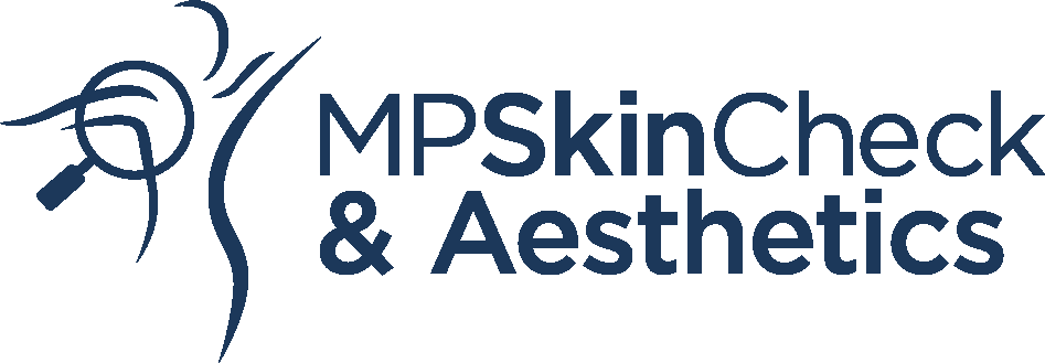
Cryotherapy is a treatment where extreme cold is used to destroy abnormal skin cells. Instead of burning with heat, the cold causes controlled cell damage.

It is commonly used for:

  • Precancerous lesions such as solar keratoses (sunspots)
  • Benign lesions such as seborrhoeic keratoses and skin tags
  • The treated area may look red immediately after treatment.
  • A blister may or may not form.
  • The wound then enters a scabby stage before flaking off.
  • Healing time:
    • 7–10 days for solar keratoses (sunspots)
    • Several weeks and sometimes multiple treatments for thicker lesions (e.g. seborrhoeic keratoses)
  • Apply a thin layer of moisturiser (e.g. Vaseline or Paw Paw) as soon as you get home.
  • Reapply several times daily for the first 48 hours.
  • If blisters form:
    • Keep the area clean.
    • If oozing, gently clean and cover with a temporary dressing until dry.
  • Hypopigmentation (whitening of the skin) at the treatment site is common and usually permanent.
  • Scarring is uncommon but possible with deeper freezes.

Please contact the clinic if you notice increasing pain, redness, or swelling after 3 days, as this may indicate infection.

Cryotherapy

Curettage and Cautery

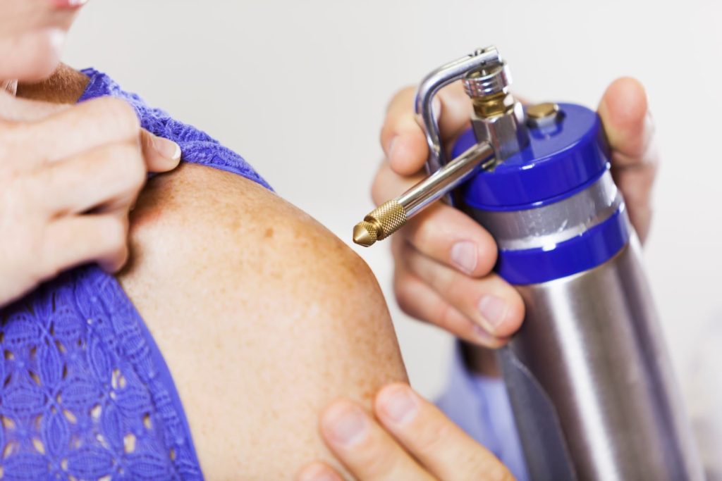
Curettage and cautery is a technique where skin cancers are removed by scraping and heat treatment. It can be a good option for certain types of skin cancer, particularly superficial lesions, and allows multiple spots to be treated in a short session.

Curretage and Cautery
  1. The area (including a small margin of healthy tissue) is numbed with local anaesthetic.
  2. The lesion and the top layer of skin are gently scraped off with a sharp instrument.
  3. The wound is then cauterised (treated with heat) using an electrical device to control bleeding.
  4. This scraping–cautery cycle is usually repeated two or three times.This both removes cancer cells and triggers an immune response to help destroy any remaining abnormal cells.
  • The wound will look like a deep graze or burn.
  • A crust will usually form, and a yellow base may appear under the wound — this is normal.
  • When the anaesthetic wears off, you may feel mild discomfort, similar to a scratch.
    This is usually relieved with paracetamol (Panadol).
    Please do not take ibuprofen (Nurofen), as it can increase bleeding risk.
  • Remove the dressing after 48 hours.
  • Wash the wound gently with water, pat dry, and apply Vaseline to keep it moist.
  • Do not stop blood-thinning medicines (unless specifically advised by your doctor).
  • If bleeding occurs:
    Apply firm pressure for 15–20 minutes.
    If you cannot stop the bleeding, contact the clinic.
  • Increasing pain, redness, swelling, or pus after a few days
  • Bleeding that won’t stop
  • Feeling unwell after treatment

After healing, the treated skin may look different:

  • A depressed scar (slightly sunken)
  • A raised scar (keloid)
  • Loss of pigment (paler skin)
  • Hyperpigmentation (darker skin)
  • Quick and efficient — multiple skin cancers can often be treated in a single session
  • Minimally invasive — no sutures required
  • Good for superficial lesions that do not need deeper excision

Excision

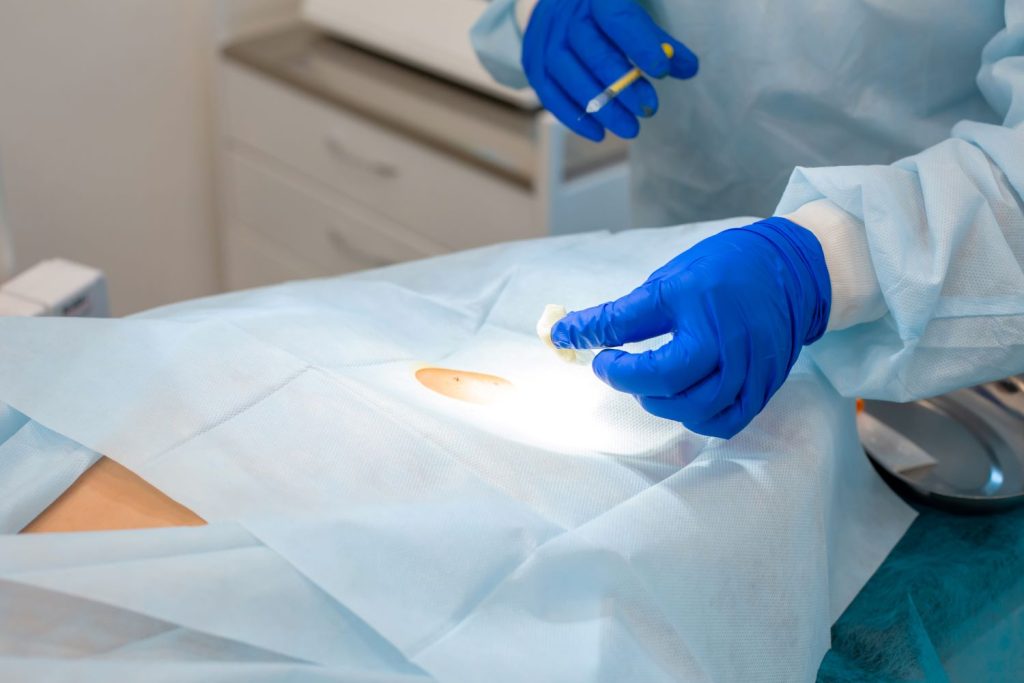
The vast majority of skin cancers can be removed under local anaesthetic with a simple elliptical excision.

  • The skin cancer is marked out, and a safety margin is drawn around the lesion.
  • Local anaesthetic is injected along the planned excision. This may sting for a few seconds but quickly takes effect.
  • The lesion is removed with a scalpel, and the wound is sutured together — usually with a mix of dissolving and non-dissolving stitches. Occasionally, only dissolving sutures are used.
  • The procedure usually takes 15–45 minutes, depending on the size and location.
  • Mild pain afterwards is common.
  • A scar will always occur after surgery.
  • Infection and bleeding risk is around 1 in 100.
  • Important: Do not stop blood thinners (e.g. aspirin, clopidogrel, warfarin, apixaban, Xarelto, Eliquis) unless specifically advised by your doctor.

Sometimes a wound cannot be closed directly because there isn’t enough loose skin. In these cases, a full-thickness skin graft may be used.

  • A small piece of skin is removed from another site (using a simple ellipse) and sewn into the new location.
  • Common donor sites: behind the ear, collarbone, or inner arm.
  • Grafts are often used for skin cancers on the nose, ears, scalp, hands, forearms, and lower legs, where direct closure is difficult.
  • Following the operation, it is essential to keep the graft clean, dry, and protected until healing occurs.

A skin flap uses tissue adjacent to the wound to close the defect.

  • This allows nearby skin with good blood supply to be repositioned over the excision site.
  • Flaps are particularly useful in areas where appearance and function matter, such as the face and nose.
  • While many smaller flaps are performed in general skin cancer practice, larger and more complex flaps are usually undertaken by plastic surgeons.

In select areas, it may be better to leave the wound open and allow it to heal naturally. This is called secondary intention healing.

  • The wound is dressed and gradually fills in from the bottom up over 4–6 weeks.
  • Common sites: creases of the nose, parts of the ear, and areas near the eye where natural healing produces good cosmetic outcomes.
  • This is a safe, low-tech option that avoids the need for grafts or flaps.
Ellipse

Efudix (5-Fluorouracil)

Efudix

Efudix for the Treatment of Solar Keratoses

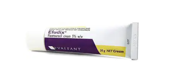
Solar keratoses (actinic keratoses) are precancerous sun-damaged lesions that appear as rough, red, scaly patches on areas like the face, ears, scalp, forearms, and hands. While most are harmless, a small number progress to squamous cell carcinoma each year (1 in 100 to 1 in 200 risk per spot per year).

Efudix is a “field treatment,” meaning it targets both visible lesions and subclinical precancerous cells in the surrounding skin. This makes it particularly effective when multiple lesions are clustered in one region.

  • Face: Apply a thin layer twice daily for ~3 weeks. Once redness/spotting occurs (usually after 1 week), reduce to once daily for the remainder.
  • Scalp, Hands, Forearms, Legs: These areas respond more slowly and often require skin preparation first: exfoliate gently for 1 week (e.g. loofah in the shower) and apply a urea-based moisturiser (e.g. Heel Balm, Ureaderm) twice daily. Then apply Efudix twice daily for up to 4 weeks.
  • Chest/Trunk: Often reacts strongly; reduce to once daily if redness becomes brisk.
  • Maximum Area: Only treat up to 500 cm² at once (roughly the size of the face, both hands, or one shin/forearm). Larger areas must be treated in stages.
If the reaction is too slow, too intense, or you are concerned, contact the clinic. Same-day reviews for Efudix reactions are available.

Efudix for the Treatment of Squamous Cell Carcinoma in situ (Bowen’s Disease)

Bowen’s disease (SCC in situ) is an early form of squamous cell carcinoma where cancerous cells are confined to the top layer of skin. Lesions usually appear as red, scaly plaques that slowly enlarge.

Efudix can be a very effective non-surgical treatment for Bowen’s disease, especially on areas where surgery might be difficult or leave large scars.

  • Apply a thin layer twice daily for 6 weeks
  • Reactions (redness, crusting, soreness) are expected and indicate the treatment is working
  • Large treatment areas may need to be staged due to the 500 cm² limit

A review is essential after treatment to ensure the lesion has resolved. If there is incomplete clearance, surgery or another therapy may be recommended.

⚠️ Contact the clinic if you have any concerns. Whilst discomfort and some redness is common, we do not expect pain that interferes with sleep, your usual activities or requires paracetamol.

Aldara (Imiquimod)

Superficial Basal Cell Cancer

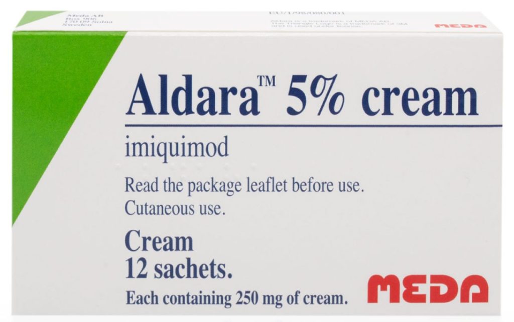
Basal cell carcinoma (BCC) is the most common and least dangerous form of skin cancer. A common subtype is superficial BCC, which often looks like a shiny pink or red patch or a dry scaly area that slowly enlarges over months or years.

Aldara works by stimulating your immune system to recognise and destroy cancer cells. The active ingredient, imiquimod, activates immune cells that then attack the abnormal skin. This process causes redness, flaking, scabbing, and sometimes soreness in the treated area — all signs that the cream is working. Very rarely, people can experience flu-like symptoms.

  • Common reactions: redness, itching, flaking, or scabbing at the site.
  • When to call us: if pain stops you from sleeping, interferes with daily activities, or requires regular pain relief.
  • Important: if reactions are severe, stop the treatment and contact the clinic. Do not try to “make up” missed doses. We can help manage troublesome reactions if they occur.
  • Apply once daily at bedtime, five days a week (usually Monday–Friday) for six weeks.
  • Wash hands and the treatment area with mild soap and dry well.
  • Apply a thin layer to the affected area plus 1 cm margin of surrounding skin.
  • Rub in gently and leave for 6–10 hours. Cotton gauze may be used to protect the area overnight.
  • Wash off thoroughly with mild soap and water the next morning.
  • Avoid eyes, lips, and nostrils.

Sachets: single-use only. Discard after opening.
Pump dispenser: replace cap after use. Store below 25 °C. Use within 4 weeks of opening. Do not freeze.

  • Avoid cosmetics, creams, or lotions on the treated area during therapy.
  • Sun protection: avoid natural and artificial UV exposure while using Aldara. Sunscreen can be applied after the treatment course is finished.
  • Aldara can rarely cause flu like symptoms, if this occurs stop treatment and notify the clinic.
Aldara

Pros & Cons of Aldara

Pros​
Cons

Join Our Mailing List